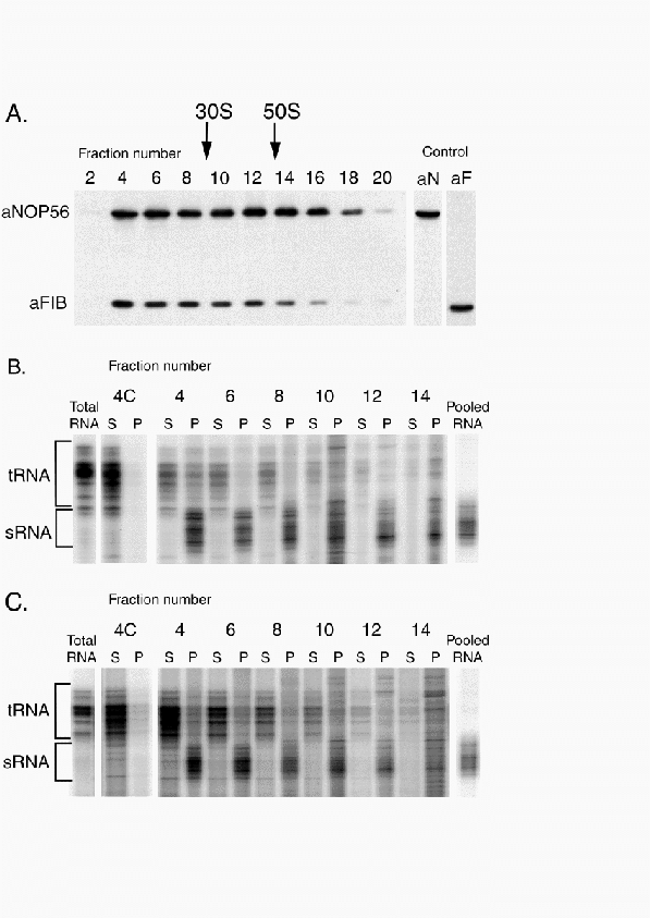
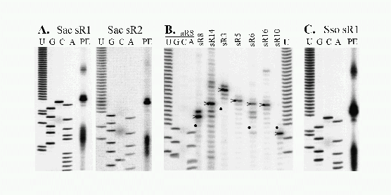
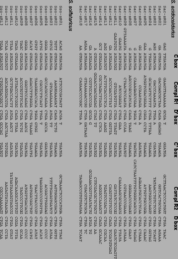
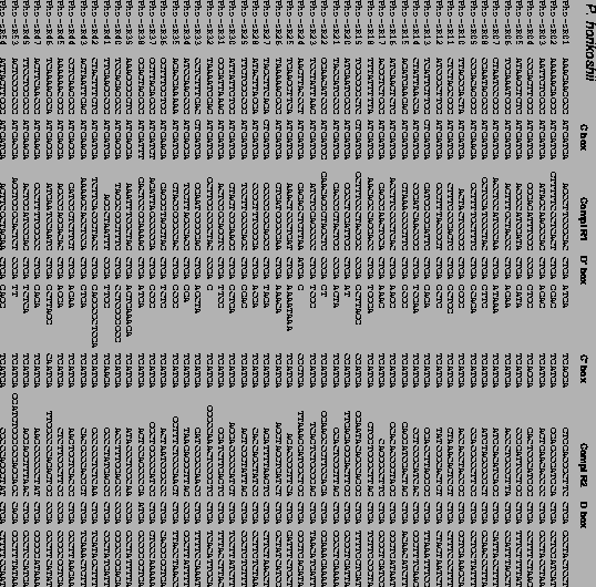
next up previous contents
Next: Bibliography Up: Small Nucleolar RNAs in Genomes.2 Previous: rRNA Methylation and Hyperthermophily

Conclusions

These findings imply that an RNA guide mechanism for directing 2'-O-ribose methylation to specific positions in rRNAs was already well established in the common ancestor of archaea and eukaryotes [Woese et al., 1990]. Neither a fibrillarin homolog nor C/D box sno-like RNAs have been described in bacteria. Therefore, it is not clear whether C/D box sRNAs were ancestral to the three surviving lineages and then either lost from or not incorporated into the bacterial lineage. Alternatively, C/D box RNAs may be a derived feature in a common ancestor of Archaea and Eukarya.


Table 5.1: Predicted Target Ribose Methylation Sites for Sulfolobus acidocaldarius sRNAs.

``Ab'' column indicates protein antibody used to co-precipitate sRNA, either $\alpha$-aFIB ``F'', or $\alpha$-aNOP56 ``N''. ``sRNA PE'' column indicates sRNAs verified to have primer extension products of the correct approximate length. ``Guide Box'' indicates the box adjacent to complementarity. ``Match/Mismatch'' indicates the number of Watson-Crick and G-U pairings versus the number of all other pairs in the guide region/target RNA duplex. ``Methyl Site Confirmed'' indicates predicted methylation sites confirmed by the primer extension pause assay [Maden et al., 1995,Kiss-Laszlo et al., 1996].

sRNA Ab sRNA Guide Box Target Site Match/ Methyl Site
    PE ?     Mismatch Confirmed?
Sac-sR1 F $\bullet$ D 16S U52 (W) 11/0 $\bullet$
Sac-sR2 F $\bullet$ D 23S C1914 11/0 $\bullet$
Sac-sR3 F $\bullet$ D 23S G2739 10/0 No
Sac-sR4 N $\bullet$ D 23S G1995 10/0 ND
Sac-sR5 F,N $\bullet$ D 16S G1056 (W)   $\bullet$
Sac-sR6 F $\bullet$   NF    
Sac-sR7 F $\bullet$ D' 23S G2649 9/0 $\bullet$
      D 23S U2692 10/0 $\bullet$
Sac-sR8 N $\bullet$ D' 23S U2972 9/1 ND
      D 23S G334 12/1 $\bullet$
Sac-sR9 F $\bullet$ D' 16S G926 8/0 ND
Sac-sR10 F $\bullet$ D' tRNA Gly-CCC C50 12/0 ND
      D 23S C2539 9/0 ND
Sac-sR11 F $\bullet$ D' 23S A2618 10/0 ND
      D 23S A724 11/2 ND
Sac-sR12 F $\bullet$ D' 23S G1114 11/0 No
      D 23S A1134 9/1 $\bullet$
Sac-sR13 N No D' 23S G385 10/1 ND
      D' 23S G2996 11/1 ND
      D 23S C2746 10/0 $\bullet$
Sac-sR14 F,N $\bullet$ D' 16S A468 12/0 No
      D tRNA Gln-UUG U34 10/0 ND
Sac-sR15 F $\bullet$   NF    
Sac-sR16 N $\bullet$   NF    
Sac-sR17 F No   NF    
Sac-sR18 N ND D' 23S G140 9/1 ND


Table 5.2: Predicted Target Ribose Methylation Sites for Sulfolobus solfataricus sRNAs.

``sRNA PE'' column indicates sRNAs verified to have primer extension products of the correct approximate length. ``Guide Box'' indicates the box adjacent to complementarity. ``Match/Mismatch'' indicates the number of Watson-Crick and G-U pairings versus the number of all other pairs in the guide region/target RNA duplex. ``Methyl Site Confirmed'' indicates predicted methylation sites confirmed by the primer extension pause assay [Maden et al., 1995,Kiss-Laszlo et al., 1996].

sRNA sRNA Guide Target Site Match/ Methyl Site
  PE? Box   Mismatch Confirmed?
Sso-sR1 $\bullet$ D' 16S U605 10/0 $\bullet$
    D' 16S U33 8/0 ND
    D 16S U52 (W) 10/0 $\bullet$
Sso-sR2 $\bullet$ D 16S G1372 10/0 ND
Sso-sR3 $\bullet$ D 16S C1490 10/0 ND
Sso-sR4 $\bullet$ D' 16S G473 8/0 ND
    D' 23S G810 8/1 ND
    D 16S C277 9/1 $\bullet$
Sso-sR5 $\bullet$ D' 23S A1183 11/0 ND
    D 23S A685 10/0 ND
Sso-sR6 $\bullet$ D' 23S G2127 9/0 ND
    D 23S G2094 10/0 $\bullet$
Sso-sR7 $\bullet$ D 16S C481 10/0 $\bullet$
    D' 23S A2425 10/0 ND
Sso-sR8 $\bullet$ D 16S U477 9/0 No
Sso-sR9 $\bullet$ D' 23S A682 11/0 ND
    D' 23 A2314 8/0 ND
    D 23S A1461 10/1 No
    D 23S A54 9/1 ND
Sso-sR10 $\bullet$ D 23S A2082 11/0 ND
Sso-sR11 ND D' tRNA Gln-CUG G18 10/0 ND
Sso-sR12 ND D 16S U1344 12/0 ND
Sso-sR13 ND D 16S G1018 (W) 11/0 ND


Figure 5.1: Glycerol gradient sedimentation of aFIB and aNOP56 containing particles present in S. acidocaldarius cell free extracts. (see legend next page)
\resizebox{!}{7.33in}{\includegraphics{figures/Arch-Figure1.ps}}


\begin{figure}% latex2html id marker 2756
\renewcommand{\baselinestretch}{1.0}
\...
... and displayed on an 8\% denaturing polyacrylamide gel
(right).
\par\end{figure}


Figure 5.2: Detection and 5' end mapping of sRNAs from S. acidocaldarius and S. solfataricus.

Primers specific for the D box guide region of Sac sR1 to sR17 were 5' end labeled with $\gamma-^{32}$P-ATP and polynucleotide kinase and used in extension reactions with total RNA (20 $\mu$g) isolated from S. acidocaldarius as template.


(A) The extension products obtained with Sac sR1 and sR2 specific oligonucleotide primers were run alongside a DNA sequence ladder generated with the same primers and Sac sR1 or sR2 cDNAs as template.


(B) The extension products obtained with Sac sRNA-specific primers and run with the DNA sequence ladder generated with Sac sR8 cDNA clone. For each extension reaction, the approximate positions of the 5' terminal nucleotide in the corresponding cDNA is indicated by a (>) beside the lane. [Note: there is an inconsistency in fragment lengths for this experiment between the Dennis lab (gel picture here) and my own primer extensions. This experiment will be repeated and the conflict resolved before submission of this manuscript.]


(C) The primer extension reaction was as in (A) except that the primer was specific to Sso sR1 and total RNA from S. solfataricus was used as template. The DNA ladder was generated using Sac sR1 primer and the Sac sR1 cDNA as template. The Sac and Sso primers differ at two internal positions but have the same 5' and 3' end location.

\resizebox{\textwidth}{!}{\includegraphics{figures/Arch-Figure_2_Primer_Ext.ps}}


Figure 5.3: Detection of methylation sites in 16S and 23S RNAs by primer extension pausing.

Primer extensions were carried out by the dNTP concentration-dependent primer extension assay as previously described [Lowe & Eddy, 1999]. RNA sequencing ladders of ribosomal RNA regions being assayed appear in left four lanes. To the right of sequencing ladders, pairs of reverse transcriptase primer extensions on the same RNA sample are shown. Odd lanes use high dNTP concentration (1.0 mM) reactions and even lanes contain low dNTP concentration (0.004 mM) reactions. 2'-O-methyl modified nucleotides are characterized by appearance of termination bands in low but not high dNTP concentration reactions. Proposed rRNA-sRNA guide duplexes appear below primer extension gels. [Note: disregard 7S guide diagram in part C. This will be removed in the final version of the manuscript].

\resizebox{\textwidth}{!}{\includegraphics{figures/Arch-Figure_3_Methylation.ps}}


Figure 5.4: Alignment of Sulfolobus acidocaldarius and Sulfolobus solfataricus sRNAs. Box features and complementary regions (Compl R#) labelled.
\rotatebox{270}{\resizebox{!}{5.0in}{\includegraphics{figures/Sulf-sno-align.eps}}}


Figure 5.5: Alignment of Methanococcus jannaschii and Archaeoglobus fulgidis sRNA predictions.
\rotatebox{270}{\resizebox{7.5in}{!}{\includegraphics{figures/Mj-Af-sno-align.eps}}}


Figure 5.6: Alignment of 25 Aeropyrum pernix sRNA predictions.
\rotatebox{270}{\resizebox{7.5in}{!}{\includegraphics{figures/Ap-sno-align.eps}}}


Figure 5.7: Alignment of 50 Pyrococcus horikoshii sRNA predictions.
\rotatebox{270}{\resizebox{\textwidth}{!}{\includegraphics{figures/Ph-sno-align.eps}}}


Figure 5.8: Alignment of 10 Pyrococcus sRNA homolog families.

Pho = Pyrococcus horikoshii, Pab = Pyrococcus abyssi, and Pfu = Pyrococcus furiosus sRNAs. Note strong conservation within box features and complementary regions, and lack of conservation in intervening and flanking sequences. Sequences are roughly aligned in flanking regions to show where conservation ends.

\rotatebox{270}{\resizebox{7.0in}{!}{\includegraphics{figures/Pyro-sno-align.eps}}}


next up previous contents
Next: Bibliography Up: Small Nucleolar RNAs in Genomes.2 Previous: rRNA Methylation and Hyperthermophily
Todd M. Lowe
2000-03-31