CMPS 161,
Winter 2010
Uliana Popov
Final Project: Multimodal visualization for neurosurgical planning
Final Project: Multimodal visualization for neurosurgical planning
IEEE Visualization Contest
MRI scan |
The Clinical questions are:
1. What is the relation between the lesion, functional areas and white matter tracts?
2. How can the lesion be accessed most safely?
Grey and white matters:
Input: .raw 3D files. Different variations of MRI scans.
.pdf file describing dimensions and voxel to world conversion matrices
MRI (Magnetic resonance imaging)
MRI is a medical imaging technique most commonly used in radiology to visualize detailed internal structure and limited function of the body. MRI provides much greater contrast between the different soft tissues of the body than computed tomography (CT) does, making it especially useful in neurological (brain), musculoskeletal, cardiovascular, and oncological (cancer) imaging.
T1 and T2
T1 scan uses a gradient echo, while T2 uses a spin echo. T1 is one of the basic types of MR contrast and is a commonly run clinical scan. T2 has long been the clinical workhorse as the spin echo sequence is less susceptible to inhomogeneities in the magnetic field. They are particularly well suited to edema as they are sensitive to water content.
DTI (Diffusion MRI)
DTI measures the diffusion of water molecules in biological tissues.
DWI (diffusion-weighted imaging)
DWI is highly sensitive to the changes occurring in the lesion.
FLAIR (Fluid Attenuated Inversion Recovery)
FLAIR is an inversion-recovery pulse sequence used to null signal from fluids. For example, it can be used in brain imaging to suppress cerebrospinal fluid (CSF) so as to bring out the periventricular hyperintense lesions, such as multiple sclerosis (MS) plaques. By carefully choosing the inversion time TI (the time between the inversion and excitation pulses), the signal from any particular tissue can be suppressed.
Functional MRI (fMRI)
fMRI measures signal changes in the brain that are due to changing neural activity.
SWI (Susceptibility weighted imaging)
SWI uses a type of contrast in magnetic resonance imaging (MRI) different from traditional spin density, T1, or T2 imaging. SWI uses a fully flow compensated, long echo, gradient echo (GRE) scan to acquire images. This method exploits the susceptibility differences between tissues and uses the phase image to detect these differences. The magnitude and phase data are combined to produce an enhanced contrast magnitude image which is exquisitely sensitive to venous blood, hemorrhage and iron storage. The imaging of venous blood with SWI is a blood-oxygen-level dependent (BOLD) technique which is why it was (and is sometimes still) referred to as BOLD venography. Due to its sensitivity to venous blood SWI is commonly used in traumatic brain injuries (TBI) and for high resolution brain venographies but has many other clinical applications.
Used a number of visualization tools to answer the clinical questions:
VTK (C++)
VTK is a set of open-source libraries for 3D computer graphics, image processing and visualization. VTK supports a wide variety of visualization algorithms including: scalar, vector, tensor, texture, and volumetric methods; and advanced modeling techniques such as: implicit modeling, polygon reduction, mesh smoothing, cutting, contouring, and Delaunay triangulation.
Used the tool to:
. parse and read the .raw files
. convert data to different file formats
. create color maps and isosurfaces
Paraview
Paraview is an open source, multi-platform data analysis and visualization application. It is built on top of the Visualization Tool Kit (VTK) libraries. It handles structured (uniform rectilinear, non-uniform rectilinear, and curvilinear grids), unstructured, polygonal, image, multi-block and AMR data types.
Used the tool to:
. visualize the data, create normals, isosurfaces
. convert data to different file formats
MedINRIA
MedINRIA is a platform containing a set of softwares. These modules have been developed by
the INRIA research team Asclepios at Sophia Antipolis, France. It has several modules: Image Viewer, DTI Track,
which contains routines for DTI processing and fiber tracking using Log-Euclidean metrics
developed at Asclepios, a Tensor Viewer module aiming at visualizing volume of tensors with
glyphs, a Registration Tool module, entirely dedicated to image registration, and registration
result evaluation, two modules specialize in the analysis of multiple scerosis lesions, and a
specific viewer for brain sulcal lines.
MedINRIA is a medical tool that allows to process and analyze a variety of MRI images. MedINRIA was initially developed for medical experts.
Used the tool to:
. slice by slice image view
. color mapping
. convert data to .dicom series exam
3D slicer
3D Slicer (Slicer) is a free, open source software package for scientific visualization and image analysis. Slicer is used in a variety of medical applications, including autism, multiple sclerosis, systemic lupus erythematosus, prostate cancer, schizophrenia, orthopedic biomechanics, COPD, cardiovascular disease and neurosurgery.
Slicer's capabilities include:
* Reading and writing DICOM images and a variety of other formats
* Interactive visualization of images, triangulated 3D surface models, and volume renderings
* Manual editing
* Fusion and co-registering of data using rigid and non-rigid algorithms
* Automatic segmentation
* Analysis and visualization of diffusion tensor imaging data
* Tracking of devices for image-guided procedures.
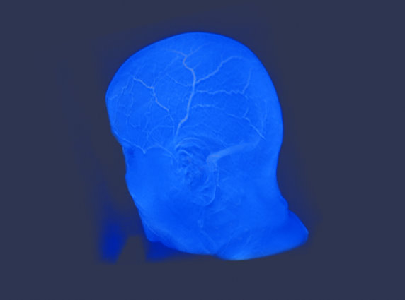
Used the tool to:
. compose a 3D volume image
. crop, color and align 3D volume with the B/W slices
What did I do?
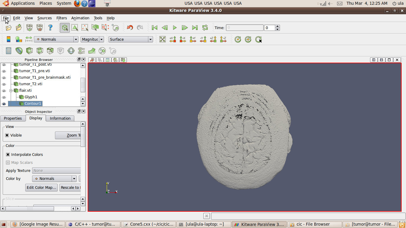
1. Read the data using VTK libraries in C++. Code.
Rendered the volumes and tried different color maps and isosurfaces.
Input format: .raw files
Output format: .vti and .vtk files
(all pictures are clickable)
|
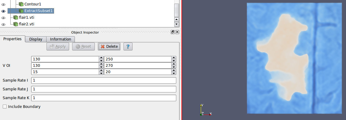
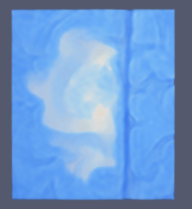
FLAIR, slice number 20 |
Tumor region |
|
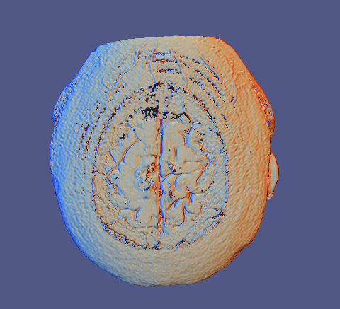
FLAIR, contour lines |
Contour lines |
|
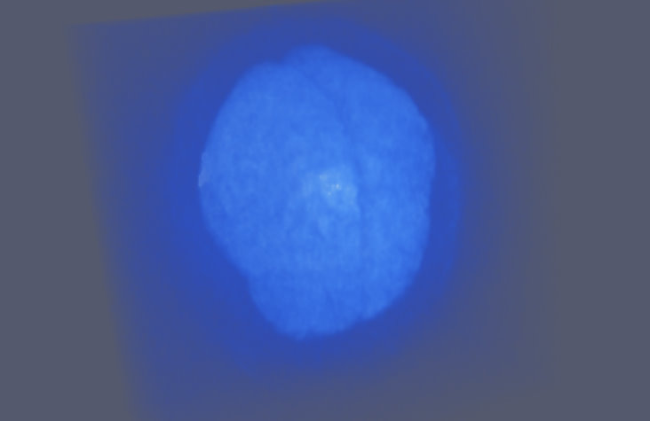
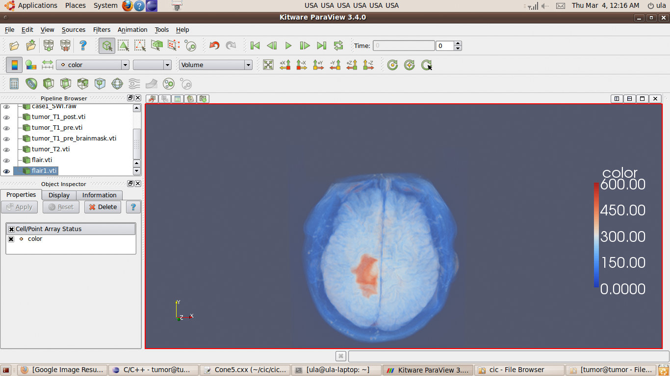
FLAIR, alfa=1: 0-600, alfa=0: >600 |
Show only 0-600 values |
It was not very successful, because I didn't know what color values correspond to the interesting regions.
I needed a more specific tool, medical oriented.
2. MedINRIA
Input format: .vtk
Output format: .dicom
Very user friendly interface. Helped a lot in understanding the data, - spatial relationship between the lesion and the risk structures.
Gives the ability to look at the data slice by slice, both axeses.
Just a reminder, one of our risk structures is:
White matter tracts:
|
|
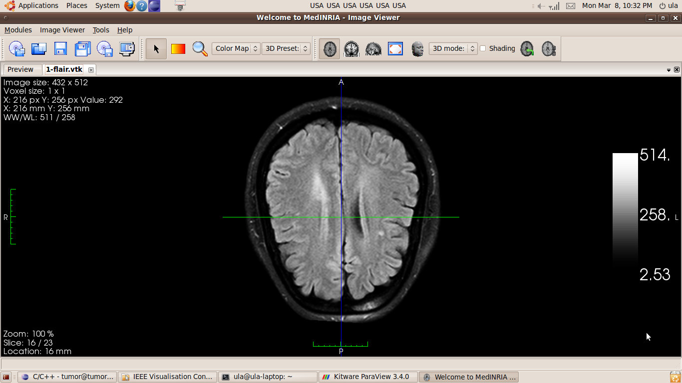
FLAIR, slice 15 out of 23.
No tumor. Tracts only. |
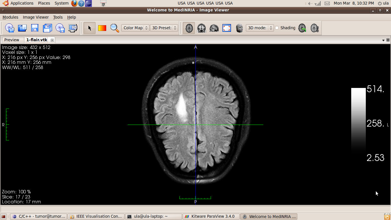
FLAIR, slice 16 out of 23.
Tumor starts to appear. |
FLAIR, slice 17 out of 23.
Tumor only. No tracts. |
According to these images, the tumor is just above the end of the tracts.
So during the surgery if we go from the top of the head, then we won't touch the white matter tracts.
We can not say anything about the grey matter though.
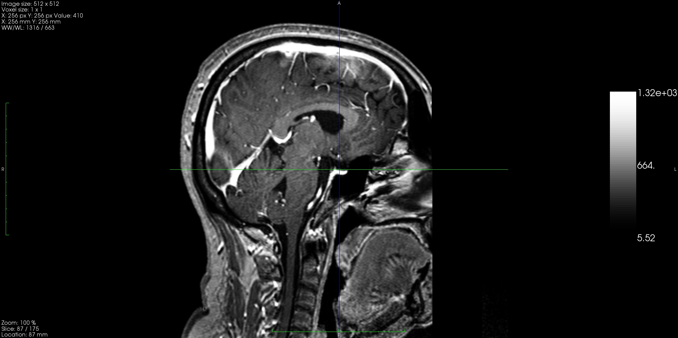
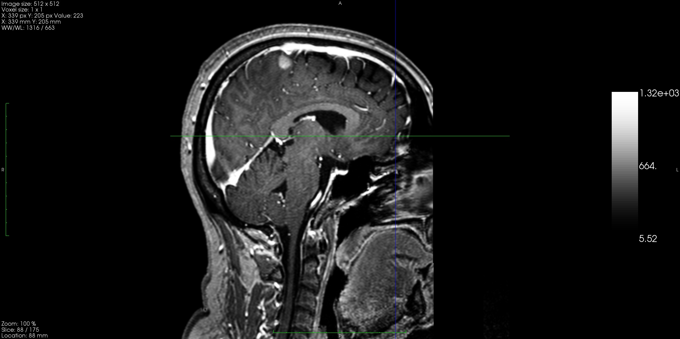
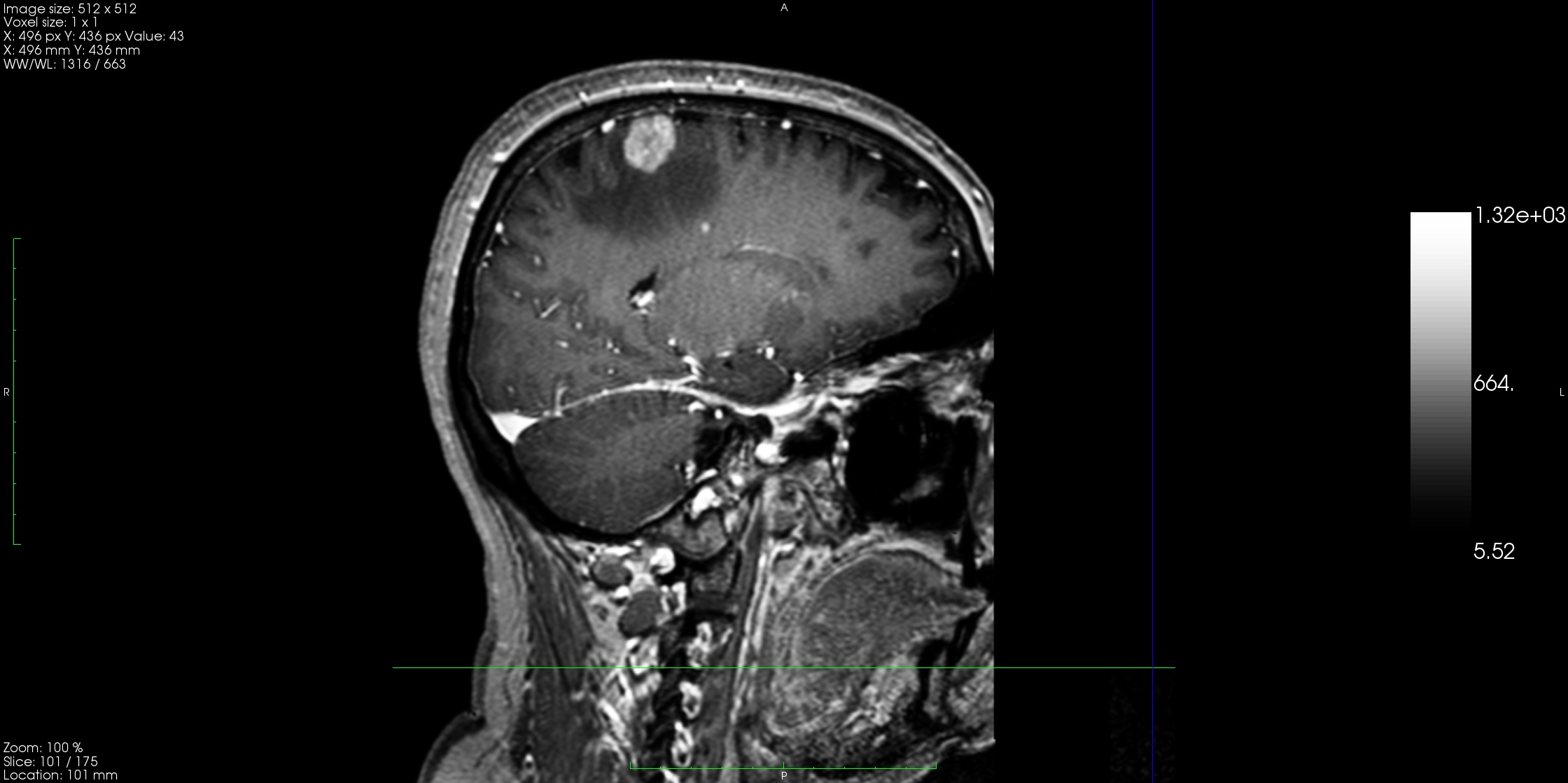
|
T1, slice 87 out of 175.
|
T1, slice 88 out of 175.
|
T1, slice 100 out of 175.
|
Here we can see that the tracts are not so close to the tracts, but we can not conclude too much about the distance to the grey matter. Does the tumor actually touches the grey matter?
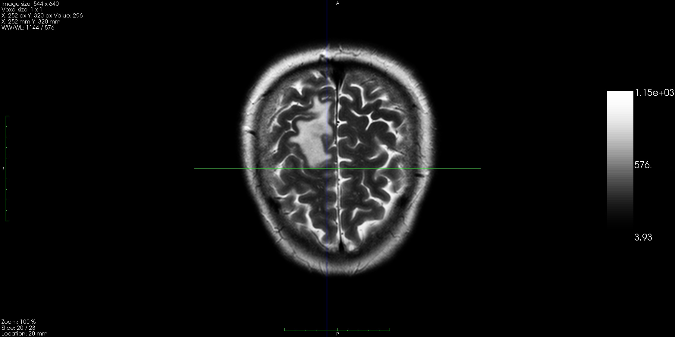
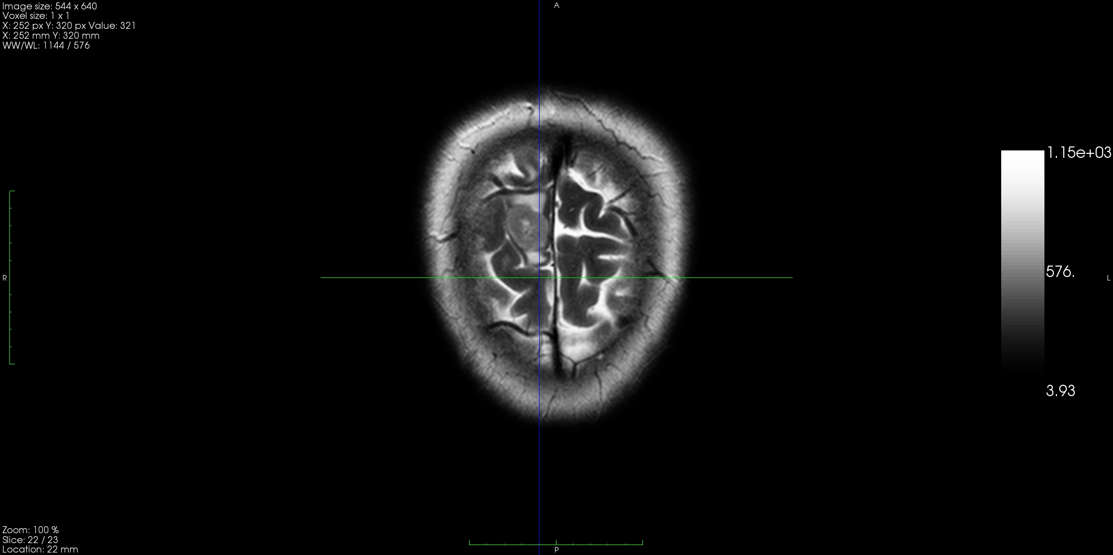
|
T2, slice 20 out of 23.
|
T2, slice 21 out of 23.
|
T2, slice 22 out of 23.
|
The question about the grey matter still anunswered. We have to find a different way to look at the images.
I tried a flow color map.
|
Here we can see better the layout of grey matter.
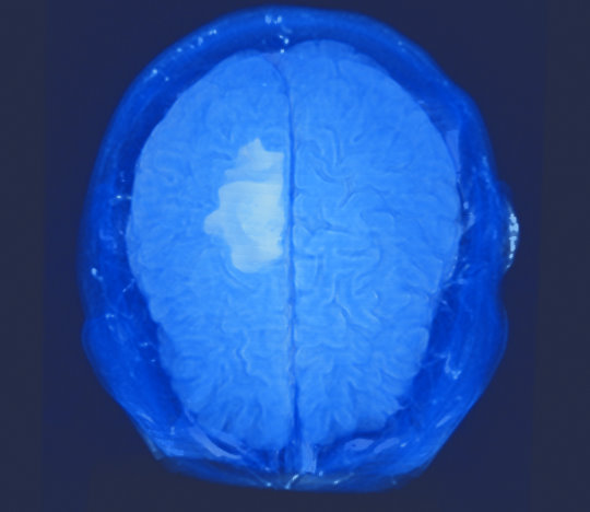
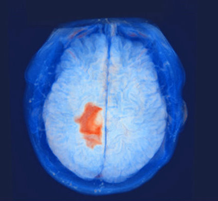
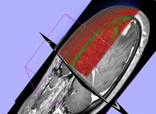
3. 3D slicer
Grey matter is green, white matter and tumor - red.
|
Finally, we can clearly see that the grey matter is already affected/damaged. So we will make the cut directly above the tumor.
A neirosurgeon confirmed my results.
Future Work
1. To map different functional areas of grey matter to the 3D volume constructed of MRI scan.
|
2. Be able to answer more specific questions, like:
- How close is the tumor located to vital functional areas, such as the visual-, language- or motor-system?
- What is the distance between the tumor and the pyramidal tract (motor), the arcuate fasciculus (associated with language processing) or the nervus opticus (vision)?
- Does the tumor infiltrate or displace any of these tracts?
- To what extend or how radical may a resection be performed?
- Which arteries or veins lie on the chosen access path?
3. To be able to handle more complicated cases.
References
1. IEEE contest web site.
http://viscontest.sdsc.edu/2010/
2. MRI:
http://en.wikipedia.org/wiki/MRI
3. MedINRIA: Medical Image Navigation and Research Tool by INRIA
www-sop.inria.fr/asclepios/software/MedINRIA/doc/.../MedINRIA.pdf
4. 3D Slicer
http://www.slicer.org/slicerWiki/index.php/Documentation-3.4
http://www.slicer.org/slicerWiki/index.php/Main_Page
5. VTK
http://en.wikipedia.org/wiki/VTK
http://www.vtk.org/doc/nightly/html/classvtkHandleWidget.html
Last modified Wednesday, 03-Mar-2010 13:44:34 PST.
| 